Breast Cancer is currently the most common cancer worldwide and more than 3300 women will be diagnosed with Breast Cancer in New Zealand this year.  
 

Early detection is our best tool to fight breast cancer

  • 3D Mammogram for clearer imagery & faster results
     
  • No Referral required if over 40 years
     
  • Mercy Breast Clinic gives you peace of mind faster by:
    • Preliminary results on the day
    • Additional screening, if requried, are available on the day
    • The ability to fast-track final results

Book A Mammogram

Book Appointment

Why go for a 3D Mammogram or Tomosynthesis

TOMO provides a 3D image of the breast producing clearer images, regardless of breast size or tissue density. With TOMO the X-Ray tube takes multiple pictures from different angles. This information is then inputted into a software program, which produces a clear three-dimensional image of the whole breast.
This provides the radiologist with multiple images, meaning they can get a clearer image of the breast tissue, rather than just the one image typically delivered by a Mammogram. This allows them to:
 
• See small lesions and other signs of breast cancer 
• Detect cancer years before you potentially have symptoms or signs of disease
• Reduce the chance of a false-positive result. The chance of having a false positive after 1 mammogram ranges from 7-12 percent, depending on your age.
 
All of this means clearer images and faster results.

More Info Book with Mercy Radiology Today

Your Mammogram with Mercy Radiology

Why Mercy Radiology & Mercy Breast Clinic?

Core Benefits of our approach include: 

  • Preliminary results on the day from our onsite radiologists
  • Additional screening if required which is available on the day if convenient
  • The ability to fast-track final results

 
This takes the pain out of waiting for your results, allowing you to get back to what matters most: Life.

Our Mercy Breast Clinic specialises in assessment and treatment of breast lumps, breast pain, breast cancer and other breast health concerns as well as breast surgical procedures. We partner with our patients throughout their journey from initial consultation, through diagnostic imaging and diagnosis; all on the same day and all in one place.

The approach to diagnosing breast problems or ruling out disease is often a complex one which requires specialist input and integration of symptoms, information with imaging results. At Mercy Breast Clinic we aim to provide you with the best possible breast care through a "one stop" clinic experience where you can undergo tests (e.g. mammogram and/or ultrasound scan – the choice of tests depends on your age, breast density type and your presenting symptom) and gain a result on the same day, as well as have a biopsy arranged and/or be assessed by a breast specialist surgeon if required – all under the one roof. Results from your tests can then be correlated together and interpreted with your actual symptom to best arrive at a diagnosis and management plan


Book A Mammogram

Book Appointment

FAQ's

Do I need a doctor’s referral for an appointment?

 You do not need a doctor's referral for a routine screening mammogram after the age of 40. You will require a doctor’s referral for an ultrasound. Please phone or email to arrange a convenient time for your appointment.

What causes  breast cancer?

Cancer grows when a cell’s DNA is damaged. But why and how that DNA becomes damaged is still unknown. It could be genetic or environmental, or in most cases, a combination of two. But most patients will never know what exactly caused their cancer. However, there are established risk factors associated with breast cancer like family history with breast cancer, early menstruation (before age 12), late menopause (after age 55), dense breast, race, use of hormone replacement therapies and alcohol use.

Risk factors are not a sure indication that you will develop breast cancer. For more information, please refer to the Breast Cancer Foundation NZ website.

Types of breast cancer?

Not all breast cancers are the same. Invasive ductal carcinoma is the most common type, representing around 70-80 percent of all breast cancers. Other types include ductal carcinoma, inflammatory breast cancer and metastatic breast cancer, which is also classified as Stage 4 breast cancer.

Breast Cancer Foundation NZ website.

How is breast cancer diagnosed and treated?

If you notice something unusual or change in your breast, or a mammogram shows something suspicious, doctors often use additional tests to find or diagnose breast cancer. Often times you will be referred to a breast specialist as these doctors are experts in diagnosing breast problems.

Imaging
Diagnostic mammogram, breast ultrasound, and sometimes Magnetic resonance imaging (MRI) scan are used to make detailed pictures of areas inside the breast.

Biopsy
This is a test that removes tissue or fluid sample from your breast. This sample is sent to the laboratory for analysis to accurately diagnose breast cancer.

Treatment
The most common form of treatment for breast cancer is surgery. An operation where doctors cut out the cancer tissue. Treatment for breast cancer can be given both locally (surgery and radiation therapy) and systematically (chemotherapy, biological therapy, hormone therapy).

Breast Cancer Foundation NZ website.

How long will my mammogram take?

It’s a good idea to allow 30min – 1 hour. Unexpected delays can occur but if there is a delay our reception team will let you know as soon as possible.

What should I wear?

Wear comfortable clothing and we suggest it is best to wear a skirt or trousers rather than a dress.

Why a mammogram?

A mammogram is still the best procedure for detecting small lumps that can’t be felt by hand. It uses low-dose x-rays to create images of the breast tissue to reveal any abnormalities or problem areas.

What age should I start getting mammograms?

At Mercy Breast Clinic, we recommend that women aged 40-50 get a mammogram every year and women over 50, every second year if there are no significant risk factors. If you have a higher risk of developing breast cancer, speak to us about when you should start having regular mammograms.

What happens during the mammogram procedure?

Firstly, we’ll ask you to undress above the waist and provide you with a wrap to put on. Next, one of our specialists will ready the machine’s plates and position your breasts up onto the plates. The upper plate will then be lowered, compressing your breasts for a short period of time while our specialist takes x-ray pictures. You may feel slight discomfort when your breasts are flattened between the plates – if it becomes too unbearable please tell us. The entire procedure will take approximately 20minutes – with the actual breast compression lasting only a few minutes. We’ll have results ready to discuss with you immediately after your mammogram.

What are the adverse effects of screening mammography?

With modern equipment and technology, the radiation dose used for your mammogram is very low. The benefits of finding and treating a breast cancer early far outweigh any risk from the x-rays. Very occasionally bruising or splitting of the skin occurs. Implant rupture has been reported however the risk is very low. Your mammographer will discuss this with you if you have implants and ask you to sign a consent to have the mammogram.

What should I do or tell the specialist before my mammogram?

Many women schedule their mammograms around their period cycle. The days leading up to your period, or during your flow, can make your breasts more tender. If you are experiencing or have experienced any breast changes or problems please talk to us about these before your mammogram. It’s also important that you inform us about any medical history that could affect your breast cancer risk—such as breast cancer in your family or hormone risk, or if you are pregnant or breastfeeding.

What is Tomosynthesis/3D Mammogram?

This is a technique that produces 3D imaging of the breast. This technology has been proven to increase the breast cancer detection rate by up to 43% as well as reduces the recall rate and any unnecessary additional testing or biopsies. There is an additional charge for tomosynthesis and this is performed in conjunction with the standard 2D mammogram. To make an informed decision, speak with the mammographer when you go into have your mammogram. Learn more about Tomosynthesis.

How common is breast cancer?

Breast cancer is the most common form of cancer in women. Early detection and treatment of breast cancer significantly improves a woman’s chance of survival.

What if I have an abnormal result?

If an area in your mammogram needs further investigation you will be contacted for another appointment with us. This appointment may involve more mammogram images, ultrasound, or a biopsy.

Size Matters: Detecting breast cancers when they are small and treatable is proven to reduce deaths from breast cancer

It is important because the smaller the tumour, the less likely it is to have spread beyond the breast and the better the chance for successful treatment. Small tumours are generally classified as two centimetres (20mm) or less.

The goal of screening is to identify cancers when they are at curable stage. The prospect for cure depends in turn on the extent to which the cancer has spread at diagnosis. It is well established that the highest cure rates are achieved in women with small, lymph node-negative breast cancers . A negative lymph node biopsy means the cancer has not spread.

Given the relationship between tumour size and survival, and given the conventional model of breast cancer spread, it is logical that the principal goal of early detection should be to identify cancers when they are small and node-negative.

3D mammography can help find breast cancer at an early stage, when treatment is most likely to be successful. A mammogram can often find breast changes that could be cancer years before physical symptoms develop.

Watch video here

Read More

Mercy Radiology & Southern Cross Health Insurance breast screening & treatment pilot

You may be eligible for an individualised and fast-tracked breast screening and diagnostic service from Southern Cross Health Insurance and Mercy Radiology

The eligibility criteria are:
1. Must be between the ages of 40 – 85 inclusive
2. Must not have had previous breast cancer
3. Must be female
4. Southern Cross Health Insurance Member

Mercy Radiology is partnering with Southern Cross Health Insurance to pilot a customised breast screening and diagnostic service. The pilot aims to recommend the best imaging for you personally by using your breast density and the clinically validated Tyrer-Cuzick risk questionnaire.

Can shorten your time to diagnosis from 6 weeks to 1.

Personalised imaging will reduce your time to diagnosis and improve the chances of finding cancers early (before they get a chance to grow). This pilot enables our specialist Radiologists to request additional imaging, if you require it, at the time of your first visit. This replaces the traditional requirement to send the initial results to your GP, who may refer you on to a specialist, who may then refer you on for additional imaging.

No matter your history, breast density, or questionnaire outcome, you will be booked for a 3D mammography (tomosynthesis) which has been shown to increase the detection of invasive cancers by 40% compared to the traditional 2D mammogram.

Other services that may be included, depending on your personal needs, include breast ultrasound, MRI or biopsy if required. All services will be covered by the one set price if they occur within 30 days of your initial appointment.

Cost:

This cost may be entirely covered by Southern Cross Health Insurance or there may be a small co-payment depending on your policy. Specialist consults and on-going management of any concerns can also be fast tracked for you, but these services will be charged separately to the pilot.

The cost is set at the beginning of your journey. If you have a shortfall, you will know exactly how much you will need to pay regardless of if you require any of the additional services included in this pilot. 

Consent: The Privacy Act 2020 and Health Information Privacy Code 2020 regulate how we can collect, use, hold, disclose, access, correct, manage, and dispose of your personal information. As per this code, we are required to manage your information safely and with respect. By taking part in this pilot, you consent to Mercy Radiology collecting and generating data and information that relates specifically to your episodes of care. We will share this information with your referring clinical team members. We will also utilise this information for our own quality improvement, research, and audit purposes. We may share anonymised and aggregated (i.e., non-identifiable) information with third-party organisations for research, quality improvement and service development purposes.
 

Watch video here

Read More

3D mammography/tomosynthesis: Imaging with more clarity for better cancer detection

It’s a piece of equipment that gives women a significantly better chance of detecting breast cancer: the 3D mammogram or digital breast tomosynthesis.

According to a large Swedish study, the Malmo study involving 15000 women screened over a 5 year period, and using the same tomosynthesis machines as those at Mercy Radiology, it was found that breast tomosynthesis detected over a third more breast cancers (34%) than traditional 2D mammography.

The difference between the 2D technology and the 3D mammography is compelling, says Dr Sugania Reddy, lead radiologist for breast imaging at Mercy Radiology. The 2D mammogram typically produces four images (two for each breast). Tomosynthesis produces multiple images of the breast depending on the size of the breast(somewhere between 60-80 images), scanning at a much more detailed level to build up 3D representations. While 2D mammograms still find many breast cancers, using tomosynthesis over the last few years at Mercy Radiology has allowed us to detect some breast cancers that could only be seen only 3D tomosynthesis. 


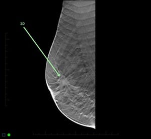
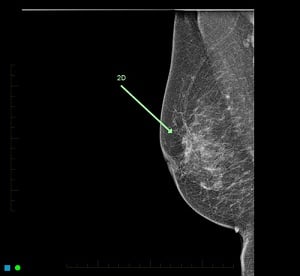
A breast cancer clearly detected on tomosynthesis (3D) and poorly on the 2D mammogram

“There’s another big difference,” says Reddy, “in that the 2D images are prone to overlapping of a breast cancer by normal breast tissue. On a 2D mammogram, the fatty tissue of the breast appears dark and fibro-glandular tissue appears as white. Unfortunately, most cancers are also white and occur in the white fibro-glandular tissue – meaning they are much harder to detect. Tomosynthesis prevents this overlap so that the cancer can be seen separately from the normal breast tissue.

A further benefit is that 3D technology also reduces patients’ recall rates. 2D technology can sometimes cause overlapping normal breast tissue on the mammogram to look like a cancer. This is reduced with tomosynthesis and therefore simply causes much less anxiety by preventing recalling a patient for extra views to confirm overlap of normal breast tissue.

Watch video here


For more information, please visit website of Breast Cancer Foundation.

Read More

Why does it matter if my breasts are dense?

Breast contains fibrous glandular, and fatty tissue.  Some women have more fat than breast tissue, while other have more breast tissue than fat.  Breast that have higher proportion of the glandular tissue are described as “dense”.

Only a mammogram can show if a woman has dense breast.  The radiologist who looks at your mammogram classifies breast composition into one of four categories of increasing density: predominantly fatty, scattered fibroglandular tissue, heterogeneously dense or extremely dense.  If you were told that you have dense breast, it means that you have either “heterogeneously dense” (C) or extremely dense” (D) breast.

The presence of dense tissue may make it more difficult to detect abnormalities in the breast and may also be associated with an increase risk of breast cancer.  Dense tissue appears white on a mammogram.  Since lumps, cancerous and non-cancerous, also appear white, they can be obscured on the mammogram.

If you have dense breasts, please talk to your doctor.  Together you can decide if supplemental screening exams can benefit you, like using tomosynthesis (as known as 3D mammogram), breast ultrasound, and MRI.

For more information, please visit website of Breast Cancer Foundation.

Watch video here

Read More

Why regular breast exam is important

Dr Benson-Cooper, an expert radiologist in breast imaging at Mercy Radiology / Mercy Breast Clinic, says mammography screening contributes to a 35 per cent reduction in breast cancer death rates, making check-ups all the more important.

Breast cancer is the leading form of cancer among women – and it kills. Every year over 3000 are diagnosed with breast cancer – primarily women over 45. Over 600 die from the disease every year.

But, when diagnosed early as part of a regular mammogram programme, the 10-year survival rate is 92 per cent.

“Breast cancer is a very serious problem in our community and can lead onto devastating outcomes. In New Zealand, 90-95 per cent of women diagnosed with breast cancer have no family history. So booking in for an annual screening mammogram is essential from the age of 40. ​After 55, if there are no risk factors, a two-yearly mammogram can be done,” Dr Benson-Cooper says. “We also recommend annual breast examinations by a GP.”

Watch video here

For more information, please visit website of Breast Cancer Foundation.

Read More

Your 1st mammogram: It will be a lot easier than you thought it would be.

Sonya Smyth, is a lead technician in Mercy Radiology with a wealth of knowledge and experience in mammography acquired over 30 years.

“If it’s your first time, you should know that it is a pretty straight forward procedure, and it’s probably not what you expect it to be.  You've heard stories about how a painful mammograms can be, and I would say that everyone who has had a first mammogram at Mercy Radiology has gone away saying “that wasn't as bad as I thought it would be”, Sonya says.   

If you are unsure of what happens, here’s a quick run-through:

  1. You need to book your appointment.  An online booking facility is available at www.radiology.co.nz or you can call our Customer Care Centre to make a phone booking.

  2. When you present for your appointment, the mammographer will explain  the procedure to you and ask you a few questions about your family history, personal breast history, if you are on HRT(Hormone Replacement Therapy), and other relevant information

  3. As for the actual mammogram, the mammographer will take 4 views of your breasts, 2 of each side, with images taken from the top and side.

  4. If you decide to have the tomosynthesis/3D mammography, the only difference you will notice is that we compress the breast a little bit longer while we do an additional scan of the breasts. This produces a more comprehensive set of images that our radiologists can scroll through to give them more information of your breast tissue.

  5. Once the mammogram is done, we ask you to wait for a few minutes while our radiologist views your images.  Sometimes we may come back to you for additional views or ultrasound and finally you will receive the radiologist's report.

  6. Your doctor will receive the report and we will add you to our recall system to inform you when your next mammogram is due.

Here’s another resource that will give you more information on what to expect in a mammogram https://radiology.co.nz/services/breast-imaging 

Watch video here

Read More

My imaging story

Marissa has a cancer history in her family and that’s why she carries out an active and preventive care of herself.  At mid 40's, she started getting annual mammograms. 

"This is something simple, something I owe to myself, my family and friends, and something that gives me peace of mind.  I also  know that getting regular screening tests can catch some cancers early, when they’re small, have not spread, and/so easier to treat.”

Whilst the real causes of many cancer may be unknown, she believes that there are ways within our hands to lower the risks of dying from it at an early stage or at a prime stage of your life.  “In my case, it’s about being good to yourself with healthy food choices, not missing my annual medical exams, and keeping away from unnecessary stresses”, says Marissa.

Marissa also added that she still gets a level of wariness each year that she does her mammogram because it still involves breast compression, but she found the latest technology called tomosynthesis more comfortable than the traditional mammogram.  It also helps when the technologist assisting you have gentle hands and makes you feel at ease all throughout the process.  “I had my latest exam with Mercy Radiology, and I immediately felt comforted that a radiologist was on site to read my report straight away, and within 10 minutes, I was cleared of potential cancer.  That gave me immediate peace of mind.”

Watch video here

Read More

Volpara: Providing the best quality image for dense breast

By adopting Volpara, it enables our radiologists to perform breast density assessment much quicker, and decide on what supplementary screening a patient may require. Using this tool gives reassurance both for us and our patients the deliver top quality image, better interpretation, and better outcome for the patients.


Read More